In this article, we will learn in detail about the important Sensory Organs in English. Read here to understand how they transmit information from the external environment to the brain. For more general information, you can check this Wikipedia link.
Introduction
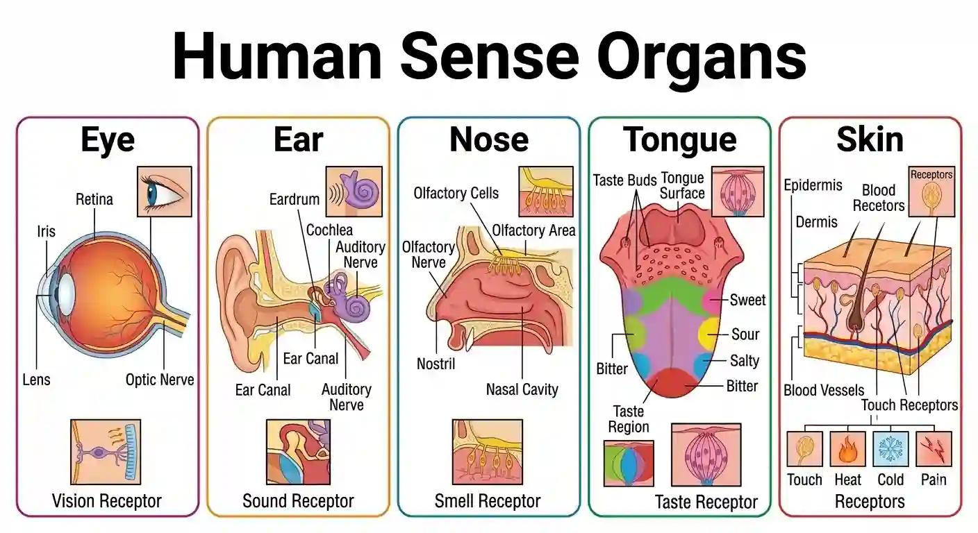
Sense Organs: “Sense” means to perceive or know, and “Organs” means the instruments or tools. Therefore, sense organs are the tools that help us perceive the world. We have five of these, which is why they are often referred to as the five senses. Alternatively, the special organs in our body that receive information from the outside world and transmit it to the brain are called “Sense Organs”. The main center that analyzes and controls all this information is our Brain. Because they introduce us to the external environment, they are aptly described as the ‘Windows of the body’.
Sense organs are the vital parts of our body that collect information from the external world and deliver it to the brain. Without them, we would not be able to comprehend what is happening around us. The scientific study of sense organs is called ‘Esthesiology’.
There are a total of five sense organs:
1. 👁️ Eye – Vision
2. 👂 Ear – Hearing and Body Balance
3. 👃 Nose – Smell (Olfaction)
4. 👅 Tongue – Taste
5. ✋ Skin – Touch/Tactile Sense
📑 Table of Contents
- Sense Organs – Introduction
- Human Eye – Structure & Diseases
- Ears – Structure & Functions
- Nose – Structure & Functions
- Tongue – Tastes & Receptors
- Skin – Layers & Functions
The scientists who first introduced the concept of the five main human senses to the world were Plato and Aristotle. They specifically emphasized the supreme importance of the sense of touch.
The scientist who first explained the crucial role nerves play in our perception of touch was Albertus Magnus.
Based on the stimuli they receive from the environment, their receptors are called as follows:
- 👁️ Eye: Receives light and provides vision – Photoreceptors
- 👂 Ear: Receives sound waves – Phonoreceptors
- 👃 Nose: Detects smell – Olfactory receptors
- 👅 Tongue: Identifies tastes – Gustatory receptors
- ✋ Skin: Perceives touch and heat/cold – Tangoreceptors (Touch) & Thermoreceptors (Temperature).
Importance of Sense Organs:
- Protection: To identify danger (e.g., immediately withdrawing your hand when you touch a hot object).
- Pleasure: To experience joy through beautiful sights, pleasant music, and delicious food.
- Communication: To talk to others and understand their feelings or expressions.
- Learning: They are the very first step to understanding the world and learning new things.
In short, sense organs act as a vital bridge between the external world and our internal world (the brain). If they do not function properly, understanding and interacting with the world becomes exceedingly difficult.
👁️ 1. Human Eye (Sensory Organs in English)
Human Eye – Structure (Sensory Organs)
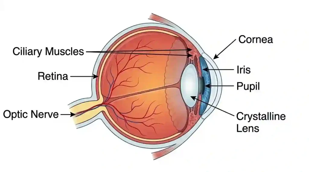
The scientific study of the eye is called Ophthalmology. The eye helps us perceive colors, shapes, sizes, and distances. This vision is possible because light enters our eyes. The eye is protected by eyelids, eyelashes, eyebrows, and lacrimal glands (tear glands). The front part of the eye is covered by a thin transparent membrane called the Conjunctiva. The eye is a spherical organ, hence called the Eyeball. The eyeball is safely situated in the eye socket of the skull. Generally, when we look at someone, only 1/6th of their eyeball is visible outside. The rest is safely embedded inside the eye socket. The human eye works like a camera. The eye is mainly protected by three layers. They are:
1. Outer Layer (Sclera)
2. Middle Layer (Choroid)
3. Inner Layer (Retina)
1. Outer Layer (Sclera)
This is the outermost tough layer that protects the eye. It is white in color and very firm. The front part of the sclera bulges out to form a transparent layer called the Cornea. During an eye donation, doctors actually extract this Cornea. It does not have a direct blood supply. At the back end of the sclera, the Optic nerve emerges and connects to the brain. The fluid secreted by the lacrimal glands keeps the conjunctiva moist and protects the eye.
2. Middle Layer (Choroid)
The choroid is generally black in color. This layer prevents internal reflection of light inside the eye. The choroid contains numerous blood vessels that supply essential nutrients and oxygen to the eye. It covers almost the entire eye except for the pupil area. The part of the choroid that surrounds the pupil is called the “Iris”. A pigment called Melanin present in the iris determines the color of our eyes (black, brown, blue, etc.). The iris consists of radial muscles and circular muscles. Behind the pupil, there is a Biconvex Lens. This lens is held in place by Ciliary muscles and Suspensory ligaments. The ciliary muscles control the biconvex lens to adjust its focal length. The small hole in the center of the iris is called the ‘Pupil’. It controls the amount of light entering the eye. The function of the iris and pupil is often compared to the diaphragm of a camera.
3. Inner Layer (Retina)
The retina is the third and innermost layer of the eye. It is the most sensitive layer. There are two types of light-sensitive photoreceptor cells in the retina: 1. Rods, 2. Cones.
Rods: These help us see in dim light (in the dark). They contain a pigment called ‘Rhodopsin’. Rhodopsin is synthesized using Vitamin A.
Cones: These help us see in bright daylight and identify colors. They contain a pigment called ‘Iodopsin’.
In the human eye, the normal ratio of rods to cones is 15:1. Nocturnal animals like owls have a higher number of rods. The part of the retina where there is absolutely no vision is called the ‘Blind spot’, and the part where the sharpest and clearest vision occurs is called the ‘Macula Lutea’ or ‘Yellow Spot’. The image formed on the retina is always real and inverted. There are two main fluid-filled chambers inside the eye: 1. Aqueous chamber 2. Vitreous chamber.
1. Aqueous chamber:
The space between the Cornea and the Lens is called the Aqueous Chamber. It is located at the front part of the eye and is filled with a thin, watery fluid called aqueous humor.
2. Vitreous chamber:
The large space behind the Lens, surrounded by the retina, is called the Vitreous Chamber. It is located in the back part of the eye and is filled with a thick, jelly-like fluid called vitreous humor.
| Feature | Aqueous Chamber | Vitreous Chamber |
|---|---|---|
| Location | Front part of the eye (between Cornea and Lens) | Back part of the eye (between Lens and Retina) |
| Fluid | Aqueous Humor | Vitreous Humor |
| Nature | Thin, Watery | Thick, Gel-like |
| Production | Continuously produced throughout life | Formed once during embryonic development |
| Main Function | Provides nutrition, regulates pressure, refracts light | Maintains the shape of the eye, protects the retina |
- Rods – Help in seeing under dim light conditions
- Cones – Help in identifying colors and seeing in bright light
The retina has two important spots:
- Blind spot – No vision is possible here
- Yellow spot – The area of the sharpest and clearest vision
☛ The yellow spot is also known as the Macula or Fovea.
● Eye Diseases and Defects Of Vision
- Night Blindness (Nyctalopia)
- Dry Eyes (Xerophthalmia)
- Short Sight (Myopia)
- Long Sight (Hypermetropia)
- Glaucoma
- Cataract
- Color Blindness
1. Night Blindness (Nyctalopia)
The inability to see objects clearly in low light or at night is called “Night Blindness”. In medical terms, it is known as Nyctalopia.
Cause: It is primarily caused by a deficiency of Vitamin-A (Retinol). Our retina contains photoreceptor cells called ‘Rods’ which help us see in the dark. Due to a lack of Vitamin-A, these rod cells fail to function properly.
Symptoms: Affected individuals can see normally in daylight, but as dusk approaches and light dims, their vision becomes blurred. They struggle in dimly lit rooms.
Prevention: Consuming a diet rich in Vitamin-A, such as carrots, spinach, papaya, eggs, and milk.
2. Dry Eyes (Xerophthalmia)
The condition where the eyes dry out due to insufficient production of tears is called “Dry Eyes” or Xerophthalmia. It can also occur due to prolonged screen time (mobile, computer, TV).
Symptoms: Burning sensation, redness, eye fatigue, and a gritty feeling in the eyes.
Prevention: Giving adequate rest to the eyes, using prescribed eye drops, and staying well-hydrated.
3. Short Sight (Myopia)
Nearby objects can be seen clearly, but distant objects appear blurry. In this defect, the image of an object is formed in front of the retina rather than on it. Myopia is corrected by using spectacles with a Concave lens.
Cause: The eyeball being too long or the lens having excessive curvature.
4. Long Sight (Hypermetropia)
Distant objects can be seen clearly, but nearby objects appear blurry. In this defect, the image is formed behind the retina. Hypermetropia is corrected by using spectacles with a Convex lens.
Cause: The eyeball being too short or the lens having insufficient curvature.
5. Glaucoma
This disease occurs when the fluid pressure inside the eye becomes abnormally high, damaging the optic nerve. An increase in Intraocular Pressure damages the Optic Nerve. The fluid in the anterior chamber (Aqueous Humor) is continuously produced and drained out through a specific pathway to maintain stable pressure. When these drainage angles are blocked or malfunction, the fluid builds up, increasing the internal eye pressure. This high pressure damages the optic nerve. If left untreated, it can lead to permanent blindness.
Symptoms:
- Eye pain
- Headaches
- Loss of vision
- Seeing halos around lights
There are primarily two types of Glaucoma:
- Open-Angle Glaucoma
- Angle-Closure Glaucoma
6. Trachoma
Trachoma is a contagious bacterial eye infection caused by the bacterium ‘Chlamydia trachomatis’. It initially affects the inner surface of the eyelids (conjunctiva). Early symptoms include mild itching, irritation, and watery eyes. If untreated, repeated infections can cause severe complications. Trachoma spreads very quickly.
Symptoms:
- Itching and burning sensation in the eyes.
- Redness of the eyes.
- Watery eyes.
- Discharge of pus or mucus from the eyes.
- Sensitivity to light (Photophobia).
7. Cataract
This condition occurs in old age when a cloudy, white layer forms over the eye’s natural lens, leading to blurred vision. It is treated surgically by implanting an artificial lens. This disease is most commonly seen in the elderly.
Symptoms:
- Cloudy or blurred vision.
- Difficulty seeing in bright light or glare.
Treatment: Surgically removing the clouded lens and replacing it with an Artificial lens.
8. Color Blindness
This is generally a hereditary condition. The inability to distinguish between certain colors is the main symptom of this disease. It occurs due to a defect or absence of Cone cells. Affected individuals typically cannot differentiate between red and green colors. The ‘Ishihara Test’ is used to diagnose this condition. Because the famous scientist John Dalton suffered from this, it is also known as ‘Daltonism’.
Cause: Malfunctioning or absence of Cones in the retina.
9. Conjunctivitis (Pink Eye)
In medical terminology, a pink eye or ‘Eye Flu’ is called Conjunctivitis. It occurs when the transparent membrane covering the eye (the conjunctiva) becomes inflamed or infected. Redness, watery discharge, itching, and eyelids sticking together are the main symptoms of conjunctivitis. Viral and bacterial eye flus are highly contagious and spread rapidly from person to person.
Vision Defects and Corrective Lenses
| Defect of Vision | Symptom / Mechanism | Corrective Lens Needed |
|---|---|---|
| ● Myopia (Short Sight) | Near objects visible, distant objects blurry. Image forms in front of the retina. | Concave Lens |
| ● Hypermetropia (Long Sight) | Distant objects visible, near objects blurry. Image forms behind the retina. | Convex Lens |
| ● Presbyopia | Age-related defect where eye muscles weaken, reducing focusing ability. | Bifocal Lens |
| ● Astigmatism | Irregular curvature of the cornea causing distorted or curved vision. | Cylindrical Lens |
📌 Parts of the Eye & Their Functions
| Part of the Eye | Function |
|---|---|
| 1. Cornea | Allows light to enter the eye |
| 2. Iris | Determines the color of the eye |
| 3. Pupil | Controls the amount of light entering |
| 4. Eye Lens | Focuses the image onto the retina |
| 5. Retina | Converts light into neural signals |
| 6. Optic Nerve | Transmits visual signals to the brain |
Questions and Answers on the Human Eye
1 Mark Questions – Answers
- Sclera
- Choroid
- Retina
2 Mark Questions – Answers
- Eyelids
- Eyelashes
- Eyebrows
- Lacrimal glands (Tear glands)
3 Mark Questions – Answers
Cause: This defect occurs when the focal length of the eye lens decreases or the length of the eyeball increases. The image of the object forms in front of the retina.
Correction: This defect can be corrected by using spectacles with a ‘Concave Lens’ of appropriate focal length.
Cause: This defect occurs due to an increase in the focal length of the eye lens or a decrease in the length of the eyeball. The image of the object forms behind the retina.
Correction: It is corrected by using spectacles equipped with a ‘Convex Lens’ of appropriate focal length.
4 Mark Questions – Answers
- Sclera – This is the tough outer layer.
- Choroid – This middle layer contains blood vessels.
- Retina – This inner layer contains light-sensitive photoreceptor cells.
- Night blindness (Nyctalopia)
- Short sight (Myopia)
- Long sight (Hypermetropia)
- Glaucoma
- Cataract
- Color blindness
- Night Blindness: This is caused by a nutritional deficiency (Vitamin-A deficiency). Due to this, the ‘Rod’ cells in the eye fail to function properly, making it difficult to see in low light or at night. It can be cured or managed with Vitamin A supplements.
- Color Blindness: This is a congenital, genetic disorder. It occurs due to a defect or absence of ‘Cone’ cells. Affected individuals cannot easily distinguish between red and green colors. Currently, there is no permanent cure for this genetic defect.
8 Mark Questions (Essay Type Answers)
Answer: The human eye is a marvelous sense organ that functions very much like a camera. It is spherical in shape and securely situated in the eye socket of the skull.
Main parts of the eye and their functions:
- Sclera: The tough outermost layer. It provides shape and protection to the eye.
- Choroid: The middle layer containing blood vessels, providing nutrients to the eye.
- Cornea: The transparent front part of the sclera. Light enters the eye primarily through this layer.
- Iris: The colored muscular ring behind the cornea. It determines the eye’s color.
- Pupil: The small opening in the center of the iris. The iris adjusts the pupil’s size depending on light intensity, controlling how much light enters.
- Eye Lens: A biconvex lens situated behind the pupil. Ciliary muscles adjust its focal length, allowing clear vision of objects at varying distances (Accommodation).
- Retina: The innermost screen-like layer. It contains photoreceptor cells called Rods and Cones.
- Optic Nerve: It converts the light signals formed on the retina into electrical impulses and transmits them to the brain.
Working Mechanism:
Light rays reflected from an object enter the eye through the cornea and the pupil. The eye lens focuses these rays to form a real, inverted image on the retina. The photoreceptors (rods and cones) in the retina get stimulated by the light and convert it into neural signals. These signals are transmitted to the brain via the optic nerve. The brain then processes and analyzes this information, enabling us to perceive the object clearly and in its upright position.
✍️ Fill in the blanks:
👂 2. Ear
Human Ear – Introduction (Sensory Organs)
● Otology: The scientific study of the ear is called Otology.
● Function: It helps us hear sounds. Ears receive vibrations (sound waves) from the air and convert them into signals the brain can understand. Ears not only help in hearing but also in regulating Body Balance.
● Bone Conduction: The process by which loud sounds reach the inner ear through the bones of the skull is called Bone Conduction.
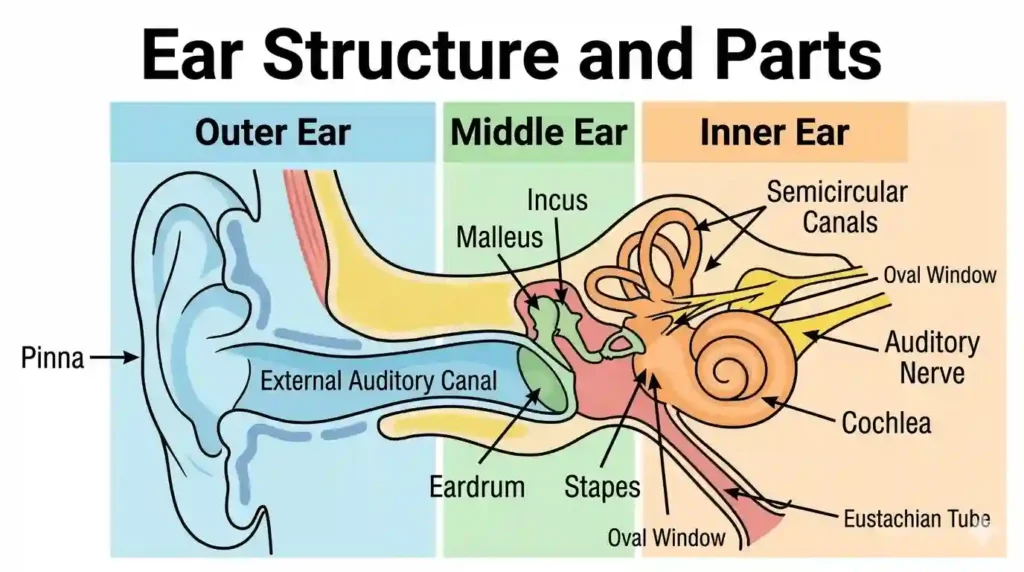
The ear has three main parts:
1. Outer Ear
2. Middle Ear
3. Inner Ear
1. Outer Ear
● Pinna: The outer visible part of the ear on either side of the head is called the Pinna or Auricle. The pinna is made of Cartilage. It opens into the Auditory Meatus (Ear canal).
● Two types of glands in the auditory meatus:
- Ceruminous glands → Secrete Ear wax
- Sebaceous glands → Secrete sebum (oil)
● These glands keep the ear canal lubricated and trap dust and debris from entering deep inside.
● Tympanic membrane / Eardrum: At the end of the auditory canal, there is a thin membrane called the Eardrum. It separates the outer ear from the middle ear. It is cone-shaped. The inner end of the eardrum is connected to the Malleus, the first bone of the middle ear.
2. Middle Ear
● The middle ear plays a crucial role in amplifying the sound vibrations that strike the eardrum. It contains a chain of three tiny bones (ossicles):
- Malleus (Hammer)
- Incus (Anvil)
- Stapes (Stirrup)
● These three bones amplify the sound intensity and transmit it to the inner ear. Eardrum + Malleus + Incus → form a chain and finally connect to the Stapes.
● The end of the middle ear is covered by a membrane called the Oval window. The middle ear connects to the inner ear through the Round window.
3. Inner Ear
● The inner ear is the most important part for hearing. It contains the Bony labyrinth, which consists of three parts: 1. Vestibule 2. Semicircular canals 3. Cochlea.
● 1. Vestibule: It has two parts:
1. Anterior part → Saccule
2. Posterior part → Utricle.
The nerve fibers from these join to form the Vestibular nerve.
● 2. Semicircular Canals: These are connected to the vestibule. They are filled with a fluid called Endolymph. The Vestibule + Semicircular canals together control body balance.
● 3. Cochlea: The cochlea has a spiral-shaped structure. It contains three canals:
- Scala vestibuli → Filled with Perilymph.
- Scala media → Filled with Endolymph.
- Scala tympani → Filled with Perilymph.
● Nerve fibers from the cochlea form the Cochlear nerve. (Vestibular nerve + Cochlear nerve = Auditory nerve).
- Pus discharge due to bacterial or fungal infections (Ear infection)
- Risk of Eardrum infections
Questions and Answers on the Ear
1 Mark Questions (Very Short Answers)
2 Mark Questions (Short Answers)
- Outer Ear
- Middle Ear
- Inner Ear
- Malleus (Hammer)
- Incus (Anvil)
- Stapes (Stirrup)
- Vestibule
- Semicircular canals
- Cochlea
3 Mark Questions
4 Mark Questions
- Pus discharge due to bacteria
- Ear infection caused by fungus
- Eardrum infections
8 Mark Questions (Essay Type Answers)
Answer: The human ear is an essential sense organ used for hearing and maintaining body balance. It is primarily divided into three parts:
● Outer Ear:
a) Pinna: It is the visible cartilaginous structure. It collects sound waves from the surroundings and directs them into the ear canal.
b) Auditory meatus: It is the canal extending from the pinna to the eardrum. It contains glands that secrete wax and oil.
● Middle Ear:
a) It begins with the Tympanic membrane (Eardrum).
b) It contains a chain of three tiny bones: Malleus, Incus, and Stapes. Sound waves striking the eardrum create vibrations. These bones amplify those vibrations and transmit them to the inner ear.
● Inner Ear:
a) It consists of the Vestibule, Semicircular canals, and Cochlea.
b) The vestibule and semicircular canals help in maintaining body balance. The spiral-shaped cochlea helps in hearing.
Working Mechanism (Hearing Process):
Sound waves collected by the pinna travel through the auditory meatus and strike the eardrum, causing it to vibrate. These vibrations are amplified tenfold by the three bones in the middle ear and reach the cochlea in the inner ear. The fluid in the cochlea creates ripples, stimulating the hair cells present there. These cells convert the sound vibrations into electrical signals (nerve impulses) and send them to the brain via the auditory nerve. When the brain processes these signals, we perceive sound.
✍️ Fill in the blanks:
👃 3. Nose
Human Nose – Internal Structure (Sensory Organs)
● Rhinology: The scientific study of the nose is called Rhinology.
● Functions of the Nose:
- Helps in respiration.
- Breathing (Inhalation and Exhalation): Before the air inhaled through the nose reaches the lungs, the tiny hairs and mucous membrane filter out dust and dirt particles. They also warm the air to match body temperature.
- Air purification.
- It perceives smell. Special cells in the nose detect odor molecules mixed in the air and send signals to the brain.
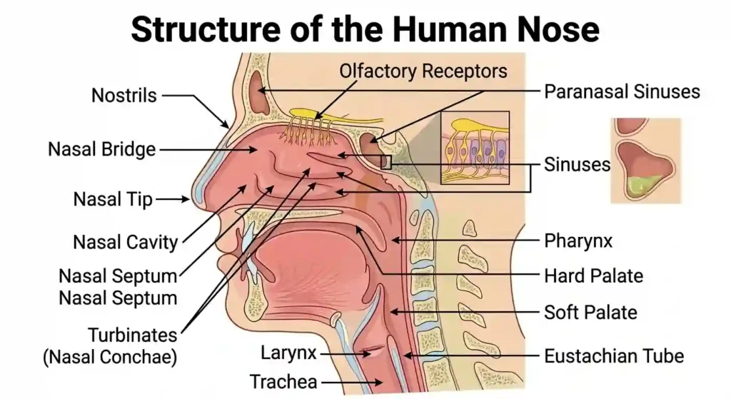
● Detecting Smell: There are olfactory receptors located in the roof of the nasal cavity. When chemical changes in the air dissolve in this mucus, the stimuli directly reach the ‘Olfactory lobes’ in the brain, allowing us to recognize the smell (fragrance or foul odor). During a cold, excess mucus is produced, covering these receptors, which is why we cannot smell properly. In dogs, this sense of smell is 40 times stronger than in humans.
● The nose has two Nostrils. These open into the Nasal cavity.
● The Nasal septum divides the nasal cavity into two halves (right and left).
● The walls of the nasal cavity are covered with a Mucous membrane and tiny hairs.
● These hairs and mucus prevent dust, microbes, and unwanted substances from entering the body.
● The mucous membrane contains Olfactory receptors. These receptors detect odor molecules, convert them into nerve impulses, and send them to the olfactory centers in the brain.
● The sense of smell in humans is comparatively lower than in many other animals.
● The receptors that perceive smell and taste are collectively known as Chemoreceptors.
📌 Structure of the Nose – Functions
| Part of the Nose | Function |
|---|---|
| 1. Nostrils | Air entry |
| 2. Nasal Cavity | Air purification / filtration |
| 3. Olfactory Receptors | Smell detection |
Questions and Answers on the Nose
1 Mark Questions (Very Short Answers)
8 Mark Questions (Essay Type Answers)
Answer: The human nose is a vital sense organ used for respiration and detecting smell (olfaction).
Structure of the Nose:
- External Nose: The visible outer part of the nose. It is made up of cartilage and bone.
- Nostrils / Nares: Two openings through which air enters.
- Nasal Septum: A thin cartilaginous layer that divides the nasal cavity into right and left halves.
- Nasal Cavity: The large empty space inside the nose. Its walls are covered with a Mucous membrane. It contains hairs that filter the air.
- Olfactory Epithelium: The roof of the nasal cavity contains special cells that detect smell. This is called the olfactory epithelium.
- Olfactory Nerve: The nerve that carries information from the olfactory receptors to the brain.
Working Mechanism (Process of Smelling):
- Chemical molecules of various smells present in the air enter the nasal cavity through the nostrils.
- These chemicals dissolve in the mucus of the nasal cavity.
- The olfactory receptors in the mucous membrane detect these chemical stimuli.
- These stimuli are converted into electrical signals and reach the olfactory centers in the brain via the Olfactory nerve.
- The brain analyzes these signals and identifies the smell we inhaled.
👅 4. Tongue
Human Tongue – Taste Receptors (Sensory Organs)
● Laryngology: The scientific study of the tongue is called Laryngology.
● Function: It helps us perceive taste. The tiny buds on the tongue (taste buds) identify sweet, astringent, sour, spicy, and bitter tastes.
● The tongue is a muscular organ. It greatly helps in tasting, mixing food with saliva for chewing, swallowing, and speaking clearly.
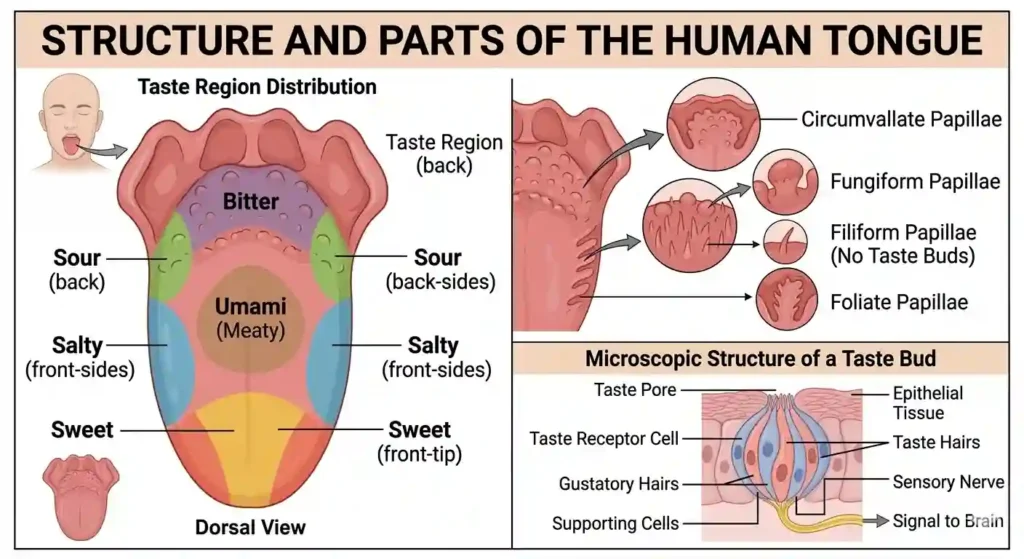
● Taste Buds: The taste buds, located in the elevated structures on the tongue called Papillae, convey the taste of the food we eat to the brain. The human tongue has about 10,000 taste buds (also known as taste receptors or gustatory calyculi). These taste buds are located in the walls of the papillae on the tongue.
● Receptors: The nerve cells that perceive taste are called ‘Gustatory receptors’.
Four Primary Tastes
- Sweet → Front part of the tongue
- Salty → Front edges of the tongue
- Sour → Sides of the tongue
- Bitter → Back part of the tongue
● Along with these, the fifth primary taste discovered by Japanese scientist Kikunae Ikeda is ‘Umami’. It is a unique savory (protein) taste found in meat, tomatoes, cheese, and soy sauce.
Types of Papillae on the tongue:
- Filiform papillae ━━━━ Scale-like structures. They do not contain taste buds.
- Fungiform papillae ━━━━ Mushroom-shaped (round) structures.
- Circumvallate papillae ━━━━ Large papillae located at the back of the tongue.
- Foliate papillae ━━━━ Ridges or bumps located on the sides of the tongue.
● Note: Except for Filiform papillae, all other papillae contain taste buds.
Questions and Answers on the Tongue
1-4 Mark Questions
- Front part of the tongue – Sweet
- Front edges of the tongue – Salty
- Back edges (sides) of the tongue – Sour
- Back part of the tongue – Bitter
8 Mark Questions (Essay Type Answers)
Answer: The tongue is a boneless, muscular sense organ. It helps in tasting, speaking, chewing, and swallowing food.
Structure of the Tongue:
There are small bump-like structures on the surface of the tongue. These are called Papillae. These papillae are mainly of four types:
- Fungiform papillae: Located on the front part of the tongue.
- Filiform papillae: Located in the middle part of the tongue (they do not contain taste buds).
- Foliate papillae: Located on the sides of the tongue.
- Circumvallate papillae: Located at the back of the tongue in an inverted ‘V’ shape.
Taste Buds:
Except for filiform papillae, all other papillae contain taste buds. Each taste bud contains taste receptor cells. These perceive tastes.
Working Mechanism (Process of Tasting):
- As we chew the food we take, it mixes and dissolves in the saliva.
- The chemicals in the dissolved food reach the taste buds through the papillae on the tongue.
- The taste receptor cells in the taste buds perceive these chemical stimuli.
- These stimuli are converted into electrical signals and reach the brain through nerves.
- When the brain processes these signals, we can recognize the taste (sweet, sour, bitter, etc.) of the substance we are eating.
✍️ Fill in the blanks:
✋ 5. Skin
Human Skin – Introduction (Sensory Organs)
● Dermatology: The scientific study of the skin is called Dermatology.
● Functions of the Skin: It perceives touch. Through the skin, we experience sensations such as heat, cold, softness, hardness, pain, and pressure. The skin also regulates body temperature and synthesizes Vitamin-D from sunlight.
● The skin is the largest organ in the human body.
● The skin is thicker on the soles of the feet and the palms of the hands compared to other parts of the body.
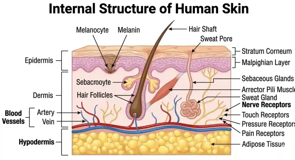
Structure of the Skin (Layers):
The skin consists of three main layers. They are:
1) Epidermis:
This is the outermost layer. It does not contain blood vessels. The pigment called ‘Melanin’, which gives color to the skin, is produced here. It protects us from the sun’s harmful UV rays. Layers within the epidermis include:
i. Stratum corneum:
- It is the outermost layer of the epidermis.
- It consists of dead cells.
- It contains a protein called Keratin.
- Keratin is also found in nails and hair.
- The cells in the stratum corneum constantly flake off as scales.
ii. Malpighian layer:
- It is the inner layer of the epidermis.
- It contains living cells.
2) Dermis:
- The dermis is the layer situated beneath the epidermis.
- Fat deposits are found below this layer.
- It contains ridges and grooves. These structures form Fingerprints.
- Fingerprints are unique to every individual, even identical twins, making them useful for identifying people.
Structures in the Dermis:
i. Hair follicles: Hair grows from the hair follicles located in the dermis.
ii. Sebaceous glands: These secrete an oily substance called Sebum. This oil prevents the skin from drying out.
3) Hypodermis:
This is the fat layer located beneath the skin. It acts as a shock absorber and a thermal insulator for the body.
Sweat glands:
- They secrete sweat. Sweat is released out through sweat pores.
- Sweat evaporates and keeps the body cool.
- Sweat glands excrete excess water, sodium chloride, and urea from the blood.
Melanin:
- The pigment responsible for the color of the skin and hair is Melanin.
- The color of the skin and hair varies depending on the amount of melanin. If melanin is abundant, the skin appears dark.
- When the skin is exposed to excess sunlight, melanin production increases, turning the skin darker. This is called Tanning.
- Melanin protects the skin from harmful Ultraviolet (UV) rays.
Skin Receptors:
- The receptors in the skin that perceive stimuli like touch, heat, and pressure are called Skin receptors.
- Receptors that detect touch are called Tactile receptors (found in higher numbers especially on fingertips and lips).
- Receptors that detect pressure are called Pacinian corpuscles.
- Receptors that detect pain in the skin are called Nociceptors.
The skin acts as a protective boundary wall for the body. However, it is susceptible to certain diseases.
- Pruritus (Itching): Occurs due to weather changes or bathing in polluted water.
- Rashes: A condition where red spots or bumps form on the skin.
- Eczema: In this disease, the skin becomes thick, turns dark gray, and flakes off.
- Pellagra: A disease caused by a deficiency of Niacin (Vitamin B₃) in the diet.
- Acne: Caused by bacteria blocking the ducts of the sebaceous glands in the skin.
- Ringworm: This is a disease caused by a fungus. Round spots form on the skin, and it becomes scaly.
- Psoriasis: In this disease, the skin sheds like scales.
- Scabies: This is a contagious disease caused by a tiny insect called Acarus / Itch mite (Scientific name: Sarcoptes scabiei). The female insect makes burrows in the skin. Sulfur-based ointments are used for prevention and cure.
Questions and Answers on the Skin
1-4 Mark Questions
- Epidermis
- Dermis
8 Mark Questions (Essay Type Answers)
Answer: The skin is the largest sense organ in the human body. It covers and protects the body and perceives touch, temperature, and pain.
Structure of the Skin:
The skin mainly consists of two layers.
- Epidermis: This is the outermost layer of the skin. It lacks blood vessels. This layer contains the pigment melanin. The epidermis protects the body by preventing the entry of microbes.
- Dermis: This is a thicker layer located below the epidermis. It contains blood vessels, nerves, sweat glands, sebaceous (oil) glands, and hair follicles.
Receptors in the Skin:
The dermis contains specialized receptors to perceive various stimuli.
- Pacinian corpuscles: Perceive Pressure.
- Meissner’s corpuscles: Perceive Touch.
- Ruffini endings: Perceive Heat.
- Krause end bulbs: Perceive Cold.
- Free nerve endings: Perceive Pain.
Working Mechanism:
When an object touches our body or when there are changes in temperature, the corresponding receptors in the skin perceive those stimuli. These stimuli are converted into electrical signals and travel to the brain via nerves. The brain then analyzes these signals, allowing us to feel the sensation (pain, heat, cold, or pressure).
📌 Match The Following
Correctly match the following fields of study with their corresponding organs:
| Study | Organ |
|---|---|
| 1. Ophthalmology | A. Skin |
| 2. Otology | B. Tongue |
| 3. Rhinology | C. Eye |
| 4. Glossology | D. Ear |
| 5. Dermatology | E. Nose |
Answers: 1-C, 2-D, 3-E, 4-B, 5-A.
📌 Organ – Field of Study
| Organ | Field of Study |
|---|---|
| ● Eye | Ophthalmology |
| ● Ear | Otology |
| ● Nose | Rhinology |
| ● Tongue | Glossology |
| ● Skin | Dermatology |
| ● Hair | Trichology |
(Note: The medical department that collectively treats diseases of the ear, nose, and throat is called ‘Otorhinolaryngology’ (ENT)).
Conclusion
Through this article, we have learned comprehensive information about Human Sensory Organs. We understood the structure and functions of these five sense organs—the eye, ear, nose, tongue, and skin—and how they transmit stimuli from the external world to the brain.
The topic of Sensory Organs is highly important for candidates preparing for Competitive Exams like UPSC, SSC, RRB, and State PSCs. Questions are frequently asked about human anatomy in the General Science and Biology section. Therefore, carefully reading and revising these Biology study notes will help you score good marks.
We hope you found this study material on Sensory Organs useful. If you have any doubts regarding this topic, please feel free to comment below.
👁️ Human Eye – Practice MCQs: